This is my personal blog.
It is also a one in all pot, thus the name "otisshairandbeatypot"
Its a pot, containing a wide range of hair and beauty product.With such as: lacewigs,customized wigs, human hairs,hair products and a wide range of beauty products.
NB;
This is an online store .
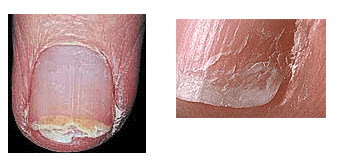
Paronychia infections of the nail fold can be caused by bacteria, fungi and some viruses. The proximal and lateral nail folds act as a barrier, or seal, between the nail plate and the surrounding tissue. If a tear or a break occurs in this seal, the bacterium can easily enter. this type of infection is characterized by pain, redness and swelling of the nail folds. People who have their hands in water for extended periods may develop this condition, and it is highly contagious. Pseudomonas bacterium
trapped between the nail
plate and the nail bed.
(“The ‘Greenies”) Pseudomonas bacterial infection can occur between the natural nail plate and the nail bed, and/or between an artificial nail coating and the natural nail plate. Many people have been led to believe that the classic ‘green’ discoloration of this type of infection is some type of mold. In actuality, mold is not a human pathogen. The discoloration is simply a by-product of the infection and is caused primarily by iron compounds. Pseudomonas thrive in moist places; it feeds off the dead tissue and bacteria in the nail plate, while the moisture levels allow it to grow. The after effects of this infection will cause the nail plate to darken and soften underneath an artificial coating. The darker the discoloration, the deeper into the nail plate layers the bacteria has traveled. If the bacteria has entered between the nail plate and the nail bed, it will cause the same discolorations and may also cause the nail plate to lift from the nail bed. Fungal Infection of the nail plate. A fungal or yeast infection which results in Onychomycosis, can invade through a tear in the proximal and lateral nail folds as well as the eponychium. This type of infection is characterized by onycholysis (nail plate separation) with evident debris under the nail plate. It normally appears white or yellowish in color, and may also change the texture and shape of the nail. The fungus digests the keratin protein of which the nail plate is comprised. As the infection progresses, organic debris accumulates under the nail plate often discoloring it. Other infectious organisms may be involved, and if left untreated, the nail plate may separate from the nail bed and crumble off. Canxidaremove can help in this case. Ringworm of the nails. Tinea Unguis, or ringworm of the nails, is characterized by nail thickening, deformity, and eventually results in nail plate loss. This is another symptom of candida yeast infection. Nail Atrophy Onychatrophia is an atrophy or wasting away of the nail plate which causes it to lose its luster, become smaller and sometimes shed entirely. Injury or disease may account for this irregularity. Ingrown Toenail Onychogryposis are claw-type nails that are characterized by a thickened nail plate and are often the result of trauma. This type of nail plate will curve inward, pinching the nail bed and sometimes require surgical intervention to relieve the pain. Vertical Split in the nail plate. Onychorrhexis are brittle nails which often split vertically, peel and/or have vertical ridges. This irregularity can be the result of heredity, the use of strong solvents in the workplace or the home, including household cleaning solutions. Although oil or paraffin treatments will re-hydrate the nail plate, one may wish to confer with a physician to rule out disease. Onychauxis Onychauxis is evidenced by over-thickening of the nail plate and may be the result of internal disorders — seek medical advice. Leuconychia Leuconychia is evident as white lines or spots in the nail plate and may be caused by tiny bubbles of air that are trapped in the nail plate layers due to trauma. This condition may be hereditary and no treatment is required as the spots will grow out with the nail plate. Beaus Lines Beau’s Lines are nails that are characterized by horizontal lines of darkened cells and linear depressions. This disorder may be caused by trauma, illness, malnutrition or any major metabolic condition, chemotherapy or other damaging event, and is the result of any interruption in the protein formation of the nail plate. Seek a physicians diagnosis. Koilonychia Koilonychia is usually caused through iron deficiency anemia. these nails show raised ridges and are thin and concave. Seek a physicians advice and treatment. Melanonychia Melanonychia are vertical pigmented bands, often described as nail ‘moles’, which usually form in the nail matrix. Seek a physicians care should you suddenly see this change in the nail plate. It could signify a malignant melanoma or lesion. Dark streaks may be a normal occurrence in dark-skinned individuals, and are fairly common. Pterygium Pterygium is the inward advance of skin over the nail plate, usually the result of trauma to the matrix due to a surgical procedure or by a deep cut to the nail plate. Pterygium results in the loss of the nail plate due to the development of scar tissue. Cortisone is used to prevent the advancement of scar tissue. Never attempt to remove pterygium -instead, consult a physician for advice and treatment.NOTE: The ‘true cuticle’ is often referred to as Pterygium. If you have pterygium, it can only be treated by a physician and should never be removed by a nail technician.
Pterygium Inversum Unguis Pterygium Inversum Unguis is an acquired condition characterized by a forward growth of the hyponychium characterized by live tissue firmly attached to the underside of the nail plate, which contains a blood supply and nerves. Possible causes are systemic, hereditary, or from an allergic reaction to acrylics or solvents. Never use force to ‘push back’ the advancing hyponychium — it is an extremely painful approach, and will result in a blood flow. Consult a physician for diagnosis and treatment. Psoriasis of the nails Psoriasis of the nails is characterized by raw, scaly skin and is sometimes confused with eczema. When it attacks the nail plate, it will leave it pitted, dry, and it will often crumble. The plate may separate from the nail bed and may also appear red, orange or brown, with red spots in the lunula. Do not attempt salon treatments on a client with Nail Psoriasis. Consult with a dermatologist for diagnosis and treatment. MMA Damage
Photo by D. Tuggle MMA Damaged Nails: D. Tuggle, owner of The Nail Academy, Jamaica, Queens, N.Y., submitted this picture of nails damaged by MMA to the BeautyTech Web Site and allowed it to be added to this page. MMA (methyl methacrylate) is a liquid monomer used for acrylic nails by some unscrupulous salons even though it is considered by and prohibited by the FDA to be a poisonous and deleterious substance. According to Dianna Bonn of Indiana, as of May 1, 1999, 23 states have banned this chemical from being used in nail salons. MMA nails are very rigid and do not bend or have the flexibility to break. When MMA does finally break, it will break the natural nail with it, causing severe nail damage. Splitting Layers & Peeling Layers Brittle Nails are characterized by a vertical splitting or separation of the nail plate layers at the distal (free) edge of the nail plate. In most cases, nail splitting and vertical ridges are characteristic of the natural aging process. This nail problem is also the result of overexposure to water and chemical solvents such as household cleaning solutions. As we age, the nail bed’s natural flow of oils and moisture is greatly reduced. This oil and moisture is the cement that holds the nail plate layers together and gives the plate its inherent flexibility. At the first signs of splitting or peeling, re-hydrate the nail plate layers with a good quality cuticle and nail oil that contains Jojoba and Vitamin E as two of the botanical oils. Jojoba oil has a very tiny molecule which can penetrate the nail plate surface, open up the layers and draw the Vitamin E in after it. The molecular structure of Vitamin E is too large to penetrate the nail plate layers or the surface layer of the skin without the benefits of Jojoba oil. Oil the nail plate and surrounding cuticle at least twice daily; more if you have your hands in water a lot. Wear gloves whenever working with household cleaning solutions, and remember: water is considered the ‘universal solvent’, and is indeed a ‘chemical’. Vertical Ridges Vertical Ridges are also characteristic of aging, although are not limited to the aged or elderly. The nail plate grows forward on the nail bed in a ‘rail and groove’ effect, much like a train rides on its’ tracks. As we age, the natural oil and moisture levels decline in the nail plate, and this rail and groove effect becomes apparent. Ridged nails will improve through re-hydration of the nail plate with twice daily applications of a good quality nail and cuticle oil containing Jojoba and Vitamin E. Hematoma A Hematoma is the result of trauma to the nail plate. It can happen from simply trapping your finger or toe in the car door to friction from improperly fitting or ‘too-tight’ shoes, to a sports related injury. A hammer does a pretty good job at causing a hematoma as well! The nail bed will bleed due to this trauma, and the blood is trapped between the nail bed and the nail plate. A hematoma may also indicate a fractured bone. Many people who participate in sports activities experience hematoma because of the constant friction from the shoes against the toenails. Hematoma may result in nail plate separation and infection because the blood can attract fungi and bacteria. If several days have passed and the blood clot becomes painful, the nail plate may require removal so the nail bed can be cleansed.
The severity of nail dysplasia is extremely variable. Nails may be small and concave, longitudinally grooved, abnormally split, pitted, softened, discolored, or brittle. Toe nails are usually less affected than finger nails.


No comments:
Post a Comment